SamanTree Medical’s Histolog® Scanner Technology Featured in 13 Publications in Special BJU International Issue on Confocal Microscopy in Urology
SamanTree Medical’s Histolog® Scanner Technology Featured in 13 Publications in Special BJU International Issue on Confocal Microscopy in Urology
Papers highlight clinical research on use of confocal microscopy for real-time tissue assessment in various urologic procedures
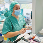
LIEGE, Belgium--(BUSINESS WIRE)--SamanTree Medical, a global leader in surgical imaging innovation, today announced that Histolog Scanner confocal microscopy technology has been featured in 13 peer-reviewed clinical papers in a special issue of BJU International, one of the world’s leading journals in urology. The special edition focuses on a growing body of evidence of the role of confocal microscopy in urologic surgery.
"Confocal microscopy is rapidly advancing as a valuable tool for tissue imaging." Dr. Ruben De Groote.
Share
“The special issue represents increasing momentum behind confocal microscopy in urologic surgery,” said Professor Greg Shaw, consultant urologic surgeon at University College London Hospitals (UCLH) and author of several studies in the special issue. “This collection, ranging from radical prostatectomy to emerging applications in additional urology procedures, demonstrates how technologies such as the Histolog Scanner can enable rapid imaging of fresh tissue.”
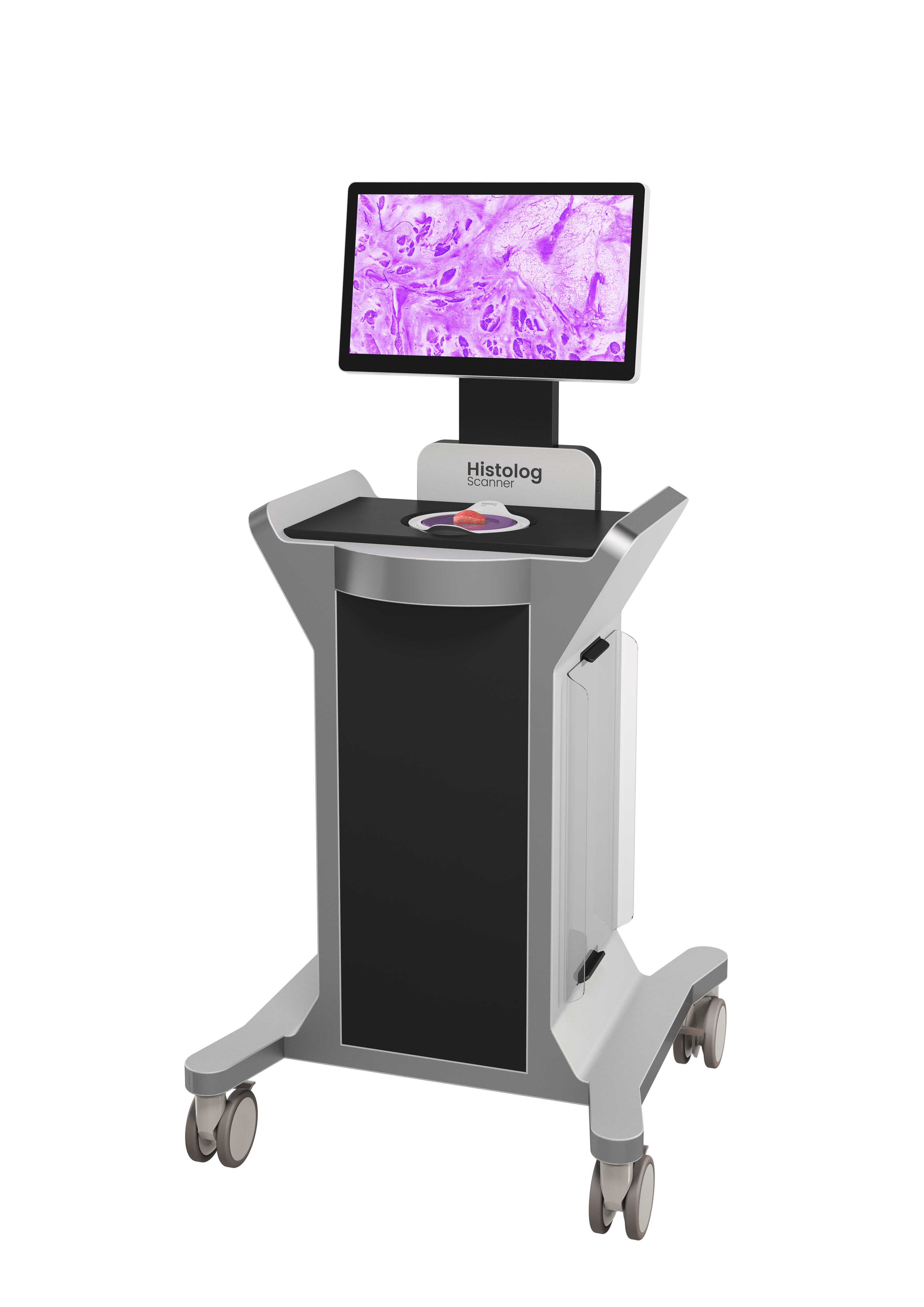
The Histolog Scanner is a confocal laser system intended to allow imaging of the internal microstructure of tissues, including, but not limited to, the identification of cells, vessels and their organization or architecture. It is the first medical imaging device using massively parallel confocal microscopy, enabling high-resolution imaging of fresh tissue surfaces within minutes without damaging the specimen.
Delphi Consensus
A central paper in the issue by Beatrici et al. reports on the results of a Delphi consensus on ex vivo fluorescence confocal microscopy in robot-assisted radical prostatectomy, which brought together international experts in urology and pathology to provide guidance on the clinical use of confocal microscopy during robot-assisted radical prostatectomy (RARP).
“Confocal microscopy is rapidly advancing as a valuable tool for tissue imaging,” said Dr. Ruben De Groote, urologist at AZORG Hospital Aalst and the organizer of the first international Delphi consensus panel on the role of confocal microscopy during RARP. “The consensus highlights the potential of rapid confocal imaging to support visualization of tissue microstructures while preserving the excised tissue for final pathology evaluation.”
Radical Prostatectomy
Several studies in the special issue evaluate the use of confocal microscopy, including the Histolog Scanner, for rapid imaging of excised tissue during RARP.
- Almeida-Magana et al. – Assessment of the feasibility of conducting a multicenter trial comparing NeuroSAFE with a novel technique based on confocal laser microscopy.
- de Angelis et al. – Clinical study evaluating intraoperative margin assessment with the Histolog Scanner during radical prostatectomy in a real-world setting.
- van Drumpt et al. – Cost-effectiveness analysis of intraoperative margin assessment strategies incorporating confocal microscopy.
- Clare et al. – Development of a histological catalogue and defined criteria for the diagnosis of prostatic adenocarcinoma and its variants using the fluorescent confocal microscope platform.
- Skok et al. – Feasibility and workflow analysis of confocal microscopy in robot-assisted prostatectomy.
- Fang et al. – Development of a deep learning model for automated interpretation of confocal microscopy images acquired during RARP.
Feasibility Studies on Emerging Applications Across Urology
Several additional papers explore the feasibility of confocal microscopy across a range of urologic procedures and diagnostic applications:
- Younis et al. – Evaluation of ex-vivo fluorescent confocal microscopy for assessment of MRI-targeted prostate biopsy specimens and quantifying workflow component times relevant to same-day implementation.
- Al-Bayati et al. – Introduction of “Co-Qual” scoring system, for assessment of prostate biopsy cores by physicians using fluorescence confocal microscopy.
- Meszes-Toth et al. – Early clinical experience of confocal microscopy for evaluation of prostate biopsy cores.
- Soputro et al. – Study evaluating confocal microscopy for reliable identification of detrusor muscle during transurethral resection of bladder tumors (TURBT).
- Daher et al. – Feasibility study evaluating confocal microscopy for rapid assessment by physicians of ureteroscopic biopsies in upper tract urothelial carcinoma.
- Zhang et al. – Study investigating the use of confocal microscopy for diagnosing penile cancer.
“What is emerging from this growing body of research is a broader recognition that surgeons benefit from having more information available while the patient is still in the physician’s office or on the operating table,” said Olivier Delporte, CEO of SamanTree Medical. “Furthermore, as these studies show early feasibility across a broader range of tissue types, SamanTree is committed to advancing research and exploring regulatory pathways to expand access to confocal microscopy technology in the U.S. and internationally.”
About SamanTree Medical
Headquartered in Liège, Belgium, SamanTree Medical is a privately held company dedicated to improving surgical precision through innovative imaging solutions. Its flagship product, the Swiss-developed Histolog Scanner, enables surgeons and pathologists to visualize fresh tissue samples in real-time. Founded in 2014 based on innovations from the École Polytechnique Fédérale de Lausanne (EPFL), a leading Swiss research institute renowned for engineering and technological advancements, SamanTree Medical is committed to improving surgical precision and outcomes by enabling real-time fresh tissue imaging. The Histolog Scanner is CE marked in the European Union and received FDA clearance in the U.S. in 2024.
U.S. Indications for Use: The Histolog® Scanner is a confocal laser system intended to allow imaging of the internal microstructure of tissues, including, but not limited to, the identification of cells, vessels and their organization or architecture.
For Important Safety Information, click here. For more information, please visit samantree.com.
Forward-Looking Statements: Certain information in these materials may include forward-looking statements regarding SamanTree Medical's products, development plans, regulatory strategy and market expectations. Any descriptions of products, indications or potential uses are for informational purposes only. Products may be used only in accordance with their approved or cleared labeling and applicable local regulations. Off-label use is not endorsed or recommended by SamanTree Medical. Actual results may differ materially. The company undertakes no obligation to update or revise these statements except as required by law.
Contacts
Media contact
SamanTree: Amy Cook | +1 925 222 5094 | amy.cook@samantree.com